Small - Rochester Advanced Dentistry")
Diagnodent
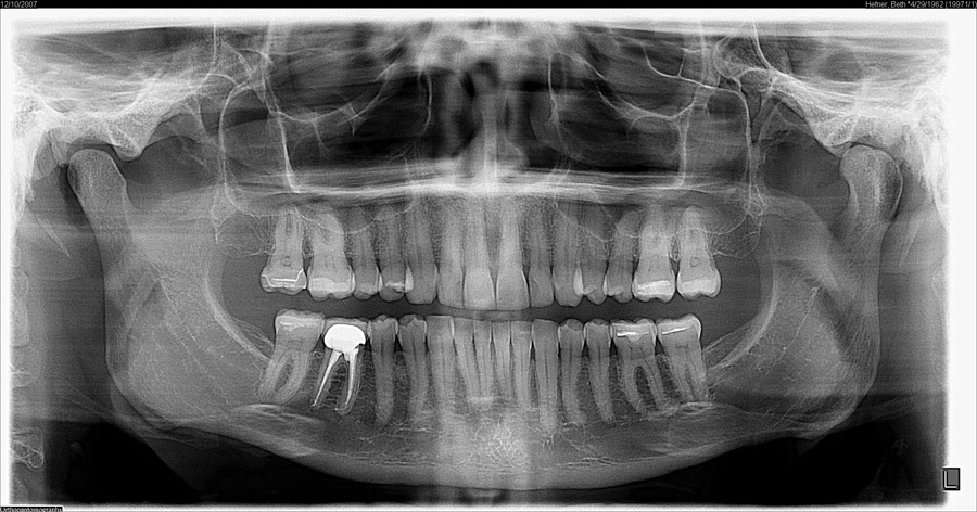
3-D Xrays
Our practice is among the first in the community to add a 3D extraoral imaging system to its office, revolutionizing patient treatment with highly detailed radiographs or X-rays. Having true-to-life 3D images produced by the device, called a CS 9300, helps Dr. Haddad provide quicker and more accurate diagnoses, improved treatment planning, and better patient care.
How are we using 3- 3-D technology in our office?
Dental Implant Placement:
3-D X-rays are instrumental in ensuring successful dental implant procedures by providing detailed imagery used for planning. While we do not perform implant surgeries in-house, we utilize these images to facilitate planning with dental professionals who will carry out your procedure. This advanced imaging allows for more predictable and streamlined implant placement. This technology is vital in enhancing the dental implant process through our collaborative efforts with dental surgeons.
Endodontic Treatment:
3-D technology has improved the accuracy of endodontic procedures by allowing our doctors to identify accessory canals, root fractures, apical abnormalities, and calcifications previously undetectable on 2-D X-rays.
Oral Surgery:
3-D X-rays are valuable in minimizing the risk of nerve damage by accurately locating the position of the inferior alveolar canal in the lower jaw and identifying other previously undetectable anatomical variabilities. While we do not perform oral surgeries in-house, this technology is crucial in aiding the dental professionals we collaborate with to avoid possible complications during and after surgical procedures.
This technology provides the advantages of high-resolution, relatively low radiation dose, and the ability to target a specific area evaluation, it is an industry-leading system designed to dramatically enhance treatment planning while delivering fast, accurate results for enhanced patient communication.
Because the CS 9300 system allows our doctors to target the exact area for exposure, dental patients are exposed to significantly less radiation when they receive an x-ray. This means other tissues around the mouth will not be unnecessarily irradiated. The system combines speed (taking X-ray images in as little as 12 seconds), image quality, and precision placement to dramatically reduce the need for retakes.
With 75-90% smaller radiation dose than conventional scans of a similar type, the CS 9300 helps us adhere to the ALARA Principle – or, ‘As Low As Reasonably Achievable’ – which we follow to ensure every precaution is taken to minimize patient exposure to radiation when obtaining the necessary diagnostic images needed for treatment planning.
With a low radiation dose and precise, crystal-clear 3D panoramic images, the CS 9300 offers unprecedented anatomical insight into specific dental regions of interest to help diagnose more accurately and treat with confidence. Benefits of the CS 9300 include:
- High-resolution images and the ability to see teeth with unprecedented detail
- Less radiation
- Comfortable positioning: the system’s open design makes exams more comfortable, with both seated and standing options to accommodate patients of all sizes
- Improved care: allowing us to perform a wider range of diagnoses with even greater accuracy
Digital Radiography
- Environmentally friendly
- Reduced exposure to radiation
- Less waiting time
- Shorter appointments
- Involved in co-diagnosis
- Better understanding of treatment
Intra Oral Camera
- Digital Pictures are taken of all areas of concern so that you are able to visualize areas of concern
Laser Cleanings
- Quickly and comfortably destroys bacteria deep in the tissue